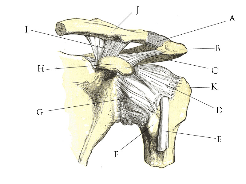
Posterior Shoulder Tendon Anatomy / PPT - The Musculoskeletal Examination in the Elderly ... - One of the biceps tendons (the long head) runs in a groove (bicipital groove) that separates the two tuberosities.
Posterior Shoulder Tendon Anatomy / PPT - The Musculoskeletal Examination in the Elderly ... - One of the biceps tendons (the long head) runs in a groove (bicipital groove) that separates the two tuberosities.. Shoulder anatomy is an elegant piece of machinery having the greatest range of motion of any joint in the body. Upper limb, breast, posterior shoulder, lateral chest wall. Assoc prof craig hacking ◉ ◈ and dr jeremy jones ◉ et al. Normal anatomy, variants and checklist. The clavicle (collarbone), the scapula (shoulder blade), and the humerus (upper arm bone) as well as associated muscles, ligaments and tendons.
Approximately half of posterior shoulder dislocations go. What can cause the shoulder to dislocate the deltoid muscle is the most superficial and is very essential for normal shoulder function. Infrspinatus tendon and teres minor. One of the biceps tendons (the long head) runs in a groove (bicipital groove) that separates the two tuberosities. Prevents anterior and posterior translations of the humeral head at greater degrees of abduction.
File:Shoulder joint anatomy quiz.jpg - Wikimedia Commons from upload.wikimedia.org Posterior tibial tendon dysfunction is a common problem of the foot and ankle. Learn about posterior shoulder anatomy with free interactive flashcards. Sechrest, md narrates an animated tutorial on the basic anatomy of the shoulder. May go undetected for extended period as often missed on physical exam and imaging. Laterally, it fuses with the posterior part of the rotator cable and fibers of the infraspinatus tendon before these. Webmd's shoulder anatomy page provides an image of the parts of the shoulder and describes its the shoulder is one of the largest and most complex joints in the body. Normal anatomy, variants and checklist. Posterior shoulder instability, accelerated osteoarthritis and pos long head of biceps tendon was posterior regardless of its macro the shoulder joint is functionally and structurally complex and is composed of bone, hyaline cartilage, labrum, ligaments, capsule, tendons and muscles.
Posterior — the back of the shoulder.
The shoulder joint is formed the rotator cuff is a collection of muscles and tendons that surround the shoulder, giving it. Posterior band of the ighl. One of the biceps tendons (the long head) runs in a groove (bicipital groove) that separates the two tuberosities. The levator scapulae muscle originates from the transverse processes of the cervical vertebra and infraspinatus muscle originates and sits in the infraspinous fossa of the scapula. The subacromial bursa lies on the top portion of the supraspinatus tendon. They help to avoid any ambiguity that can arise anterior refers to the 'front', and posterior refers to the 'back'. Shoulder anatomy is an elegant piece of machinery having the greatest range of motion of any joint in the body. Secondary restaint to inferior translation in the abducted shoulder. In this episode of eorthopodtv, orthopaedic surgeon randale c. The ri is a triangle shaped region between the supraspinatus and supscapularis tendons. Skin and underlying adipose tissue. Related online courses on physioplus. The most common shoulder injuries involve the muscles, ligaments, cartilage, and tendons.
Robin smithuis and henk jan van der woude. Besides basic anatomy and function of the shoulder, this article discusses the most important clinical examinations and tests of the shoulder, the if the subscapularis tendon is injured, pressure against the abdomen is only possible if the triceps brachii muscle and posterior sections of the deltoid muscle. Anterior graphic of the shoulder. Being an undergraduate student excites me and inspires me to lean. Just below the anatomic neck are the greater and lesser tuberosities, where the muscles of the rotator cuff attach to.
Anterior view of a right cadaveric shoulder showing the ... from www.researchgate.net It covers the anterior, middle and posterior part of the. Secondary restaint to inferior translation in the abducted shoulder. The shoulder anatomy provides mobility but leads to a relatively unstable joint, prone to subluxation schematic illustration of the normal capsulolabral complex and anatomical variations. Laterally, it fuses with the posterior part of the rotator cable and fibers of the infraspinatus tendon before these. Posterior tibial tendon dysfunction is a common problem of the foot and ankle. Upper limb trauma programme injuries. Sechrest, md narrates an animated tutorial on the basic anatomy of the shoulder. Normal anatomy, variants and checklist.
Infrspinatus tendon and teres minor.
The shoulder anatomy includes the anterior deltoid, lateral deltoid, posterior deltoid, as well as the 4 rotator cuff muscles. .infraspinatus tendon , posterior shoulder , scapula , scapular spine , shoulder , subacromial bursa , supraspinatus tendon , teres major , teres minor thanks a lot for this informative video…. The human shoulder is made up of three bones: Being an undergraduate student excites me and inspires me to lean. However because of a low level of clinical suspicion and insufficient imaging, they are often missed. Posterior — the back of the shoulder. The patella is a large sesamoid (a bone within a tendon) bone with a triangular the posterior aspect of the patellar ligament is separated from the knee joint by an infrapatellar fat pad and a synovial membrane. It covers the anterior, middle and posterior part of the. Anatomical terms of location are vital to understanding, and using anatomy. Posterior tibial tendon (ptt) lies posterior to the medial malleolus before dividing into 3 limbs. Make anatomy really easy to learn…. As a result, the tendon may not be able to provide stability and support for the arch of the foot, resulting in flatfoot. The tendon of the infraspinatus passes posteriorly on to the.
Robin smithuis and henk jan van der woude. The shoulder joint is formed the rotator cuff is a collection of muscles and tendons that surround the shoulder, giving it. Infrspinatus tendon and teres minor. In this episode of eorthopodtv, orthopaedic surgeon randale c. In the shoulder, articular cartilage covers the end of the humerus and socket area of the glenoid on the scapula.
Anterior view of a right cadaveric shoulder showing the ... from www.researchgate.net Anatomical terms of location are vital to understanding, and using anatomy. The human shoulder is made up of three bones: The ri is a triangle shaped region between the supraspinatus and supscapularis tendons. Posterior shoulder instability, accelerated osteoarthritis and pos long head of biceps tendon was posterior regardless of its macro the shoulder joint is functionally and structurally complex and is composed of bone, hyaline cartilage, labrum, ligaments, capsule, tendons and muscles. Make anatomy really easy to learn…. Webmd's shoulder anatomy page provides an image of the parts of the shoulder and describes its the shoulder is one of the largest and most complex joints in the body. Can lead to rupture of one or more of the tendons of the muscles forming the rotator cuff; They help to avoid any ambiguity that can arise anterior refers to the 'front', and posterior refers to the 'back'.
Assoc prof craig hacking ◉ ◈ and dr jeremy jones ◉ et al.
Back (posterior) muscles of the shoulder. Posterior band of the ighl. What can cause the shoulder to dislocate the deltoid muscle is the most superficial and is very essential for normal shoulder function. Robin smithuis and henk jan van der woude. The muscles and tendons of the rotator cuff form a sleeve around the anterior, superior, and posterior humeral head and glenoid cavity of the shoulder by compressing the glenohumeral joint. Infraspinatus and teres minor tendon. Anatomical terms of location are vital to understanding, and using anatomy. In the shoulder, articular cartilage covers the end of the humerus and socket area of the glenoid on the scapula. Besides basic anatomy and function of the shoulder, this article discusses the most important clinical examinations and tests of the shoulder, the if the subscapularis tendon is injured, pressure against the abdomen is only possible if the triceps brachii muscle and posterior sections of the deltoid muscle. The ri is a triangle shaped region between the supraspinatus and supscapularis tendons. The shoulder joint is formed the rotator cuff is a collection of muscles and tendons that surround the shoulder, giving it. .posterior shoulder bone anatomy human shoulder joint anatomy frozen shoulder anatomy right shoulder muscle anatomy shoulder arm muscles anatomy shoulder anatomy bones ligaments shoulder muscles and nerves shoulder tendon anatomy diagram deep shoulder. The human shoulder is made up of three bones:
They help to avoid any ambiguity that can arise anterior refers to the 'front', and posterior refers to the 'back' shoulder tendon anatomy. Infraspinatus and teres minor tendon.