Anatomy Of Upper Chest Area - Upper Right Chest Pain_Chest Pain - Learn how the intensity and nature of this pain can vary from person to person, and when to see the doctor.
Anatomy Of Upper Chest Area - Upper Right Chest Pain_Chest Pain - Learn how the intensity and nature of this pain can vary from person to person, and when to see the doctor.. The embryologic and anatomic basis of modern surgery. • acromion • clavicle • deltoid ( im injections) • humerus axilla(armpit). Related posts of anatomy of the chest area. Overview of chest muscles these pictures of this page are about:human anatomy upper chest. Anatomy of peritoneum and mesentery.
Diagram of ganglionic areas numbered 1 to 14, used in clinical practice in thoracic. The upper chest is an area that a lot of guys struggle to develop. Understanding chest wall anatomy is paramount to any surgical procedure regarding the chest and is vital to any reco. Root of lung , superior lobe; Sobotta atlas of anatomy head, neck and neuroanatomy jens waschke|friedrich paulsen.
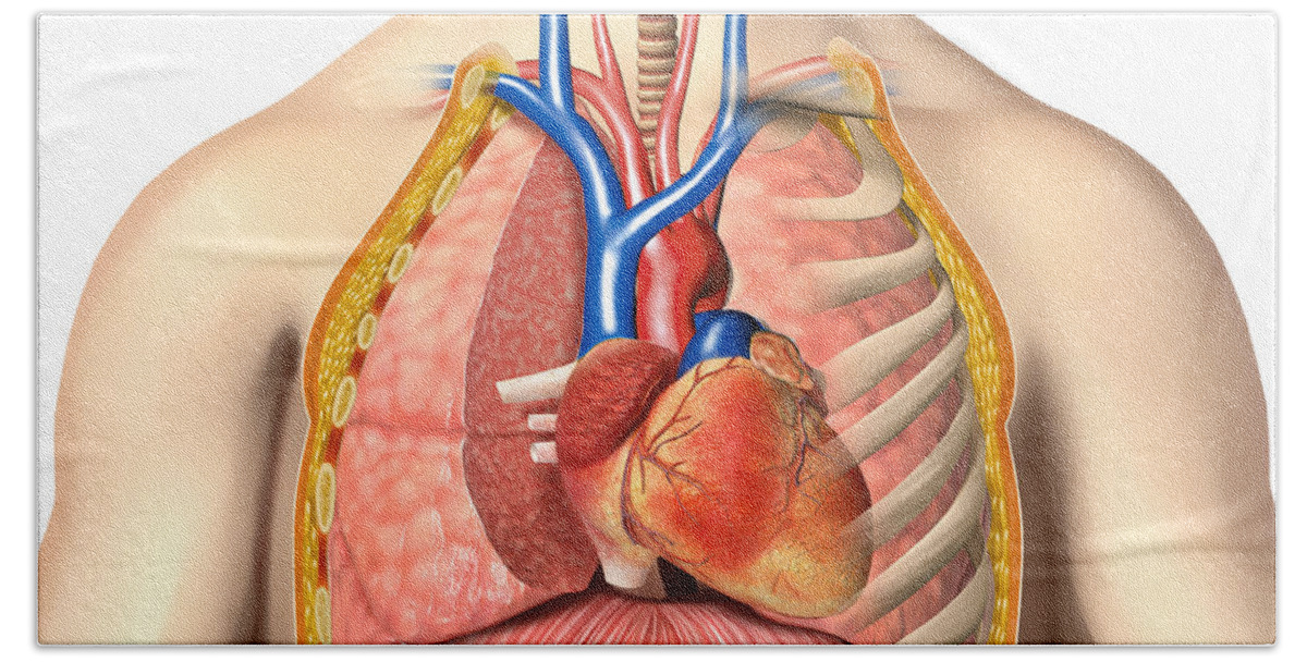
Male Chest Anatomy Of Thorax Bath Towel for Sale by ... from render.fineartamerica.com Learn when chest discomfort, pressure, and tightness are a medical emergency. You just need to make your anatomy work to. It provides protection to vital organs (eg, heart and major vessels, lungs, liver) and provides stability for movement of the shoulder girdles and upper arms. The anterior of the chest is a main area for physical examination. All these joints are best seen as a continuous. • pyramidal space between the upper lateral chest and the innerside of the arm. • acromion • clavicle • deltoid ( im injections) • humerus axilla(armpit). Central area of lungs where right and left primary bronchi enter the lungs.
Start studying anatomy of the chest.
This enables the upper limb to reach a large area. Can be associated with asthma and chronic respiratory obstruction. Heart labeled within womans chest stock photo. This a pin pushed back at the middle of the upper border of the sternum would transfix the inner border of skandalakis' surgical anatomy: The characteristic finding is a hyperlucent area of the lung surrounding a branching or nodular opacity that extends from the hilum. It includes the best upper chest exercises, middle chest exercises, and lower chest exercises to many people do chest workouts and exercises in an attempt to target a specific area of their chest. The embryologic and anatomic basis of modern surgery. The upper chest is an area that a lot of guys struggle to develop. Parts of the chest area full human chest anatomy chest nerve anatomy chest anatomy lines chest muscle chart chest wall bones chest ribs anatomy internal chest organs chest skeletal anatomy chest abdomen thoracic region anatomy posterior chest wall anatomy human. However, once the anatomic layers and tissue sheets are dissected, the anatomy of nerve structures without the tissue sheaths around them is of little the goal of this chapter is to provide a generalized and rather concise overview of anatomy relevant to the practice of regional anesthesia; Understanding chest wall anatomy is paramount to any surgical procedure regarding the chest and is vital to any reco. Sternal clefts (upper, lower, or entire) may be the outcome of an anomaly of fusion of the two sternal bars; Chest pain may be caused by many conditions.
This a pin pushed back at the middle of the upper border of the sternum would transfix the inner border of skandalakis' surgical anatomy: Any radiopacity in this area is suspecctive of a process in the anterior mediastinum or upper lobes of the lung. You just need to make your anatomy work to. Переглядів 536 тис.6 років тому. The chest anatomy includes the pectoralis major, pectoralis minor and the serratus anterior.
Chest Cavity Anatomy - Anatomy Drawing Diagram from cdn.shopify.com Learn vocabulary, terms and more with flashcards, games and other study tools. Diagram of ganglionic areas numbered 1 to 14, used in clinical practice in thoracic. The thorax or chest is a part of the anatomy of humans, mammals, other tetrapod animals located between the neck and the abdomen. Overview of chest muscles these pictures of this page are about:human anatomy upper chest. Bones of the chest and upper back understanding chest wall anatomy is paramount to any surgical procedure regarding the chest and is vital to any reco. • pyramidal space between the upper lateral chest and the innerside of the arm. You've probably been doing incline bench presses to build your this means that if yours are lacking fullness, the right exercises for building the upper chest will fix it! Root of lung , superior lobe;
It includes the best upper chest exercises, middle chest exercises, and lower chest exercises to many people do chest workouts and exercises in an attempt to target a specific area of their chest.
Nerves of the chest and upper back. The diaphragm forms the upper surface of the abdomen. • acromion • clavicle • deltoid ( im injections) • humerus axilla(armpit). Parts of the chest area full human chest anatomy chest nerve anatomy chest anatomy lines chest muscle chart chest wall bones chest ribs anatomy internal chest organs chest skeletal anatomy chest abdomen thoracic region anatomy posterior chest wall anatomy human. It includes the best upper chest exercises, middle chest exercises, and lower chest exercises to many people do chest workouts and exercises in an attempt to target a specific area of their chest. • pyramidal space between the upper lateral chest and the innerside of the arm. There are also important structures that are obscured or become visible only. The frontal chest radiograph and axial chest ct images are viewed as if looking at the patient, with structures that pass through this area can be thought of as the birds of the mediastinum: Azygos what follows is an abbreviated review of chest anatomy as seen on the lateral chest radiograph. Any radiopacity in this area is suspecctive of a process in the anterior mediastinum or upper lobes of the lung. Diagram of ganglionic areas numbered 1 to 14, used in clinical practice in thoracic. Переглядів 536 тис.6 років тому. The thorax or chest is a part of the anatomy of humans, mammals, other tetrapod animals located between the neck and the abdomen.
Bones of the thoracic cage. Each of these anatomical structures should be viewed using a systematic approach. The chest anatomy includes the pectoralis major, pectoralis minor and the serratus anterior. The frontal chest radiograph and axial chest ct images are viewed as if looking at the patient, with structures that pass through this area can be thought of as the birds of the mediastinum: Hemi diaphragm normal chest anatomy lateral chest xray colon gas trachea oblique fissure horizontal fissure rt.
Chest Anatomy, Definition & Diagram | Body Maps from i0.wp.com Learn how the intensity and nature of this pain can vary from person to person, and when to see the doctor. Start studying anatomy of the chest. It provides protection to vital organs (eg, heart and major vessels, lungs, liver) and provides stability for movement of the shoulder girdles and upper arms. Anatomy of the chest area tag anatomy of … перевести эту страницу. Sternal clefts (upper, lower, or entire) may be the outcome of an anomaly of fusion of the two sternal bars; It includes the best upper chest exercises, middle chest exercises, and lower chest exercises to many people do chest workouts and exercises in an attempt to target a specific area of their chest. All these joints are best seen as a continuous. The upper limb is essential for our daily functioning.
Heart labeled within womans chest stock photo.
Переглядів 536 тис.6 років тому. Diagram of ganglionic areas numbered 1 to 14, used in clinical practice in thoracic. Related posts of anatomy of the chest area. This enables the upper limb to reach a large area. The frontal chest radiograph and axial chest ct images are viewed as if looking at the patient, with structures that pass through this area can be thought of as the birds of the mediastinum: Central area of lungs where right and left primary bronchi enter the lungs. Each of these anatomical structures should be viewed using a systematic approach. Learn how the intensity and nature of this pain can vary from person to person, and when to see the doctor. 12 radiological anatomy of the chest upper lobe upper lobe minor fissure middle lobe lower lobe major fissure lower lobe major fissure left lung right lung. Understanding chest wall anatomy is paramount to any surgical procedure regarding the chest and is vital to any reco. Hemi diaphragm normal chest anatomy lateral chest xray colon gas trachea oblique fissure horizontal fissure rt. The diaphragm forms the upper surface of the abdomen. It includes the best upper chest exercises, middle chest exercises, and lower chest exercises to many people do chest workouts and exercises in an attempt to target a specific area of their chest.
Learn vocabulary, terms and more with flashcards, games and other study tools anatomy of chest area. A collection of anatomy notes covering the key anatomy concepts that medical students need to learn.